

Get the patient ready
Explain the exam, ask about relevant history, apply gel and position the patient so the area is reachable.
Career profile
Sonographers use ultrasound to picture inside the body, then give doctors technical records to interpret.
The picture changes whenever the transducer moves. A useful scan takes precise hands, close watch on the monitor and ongoing talk with the patient.
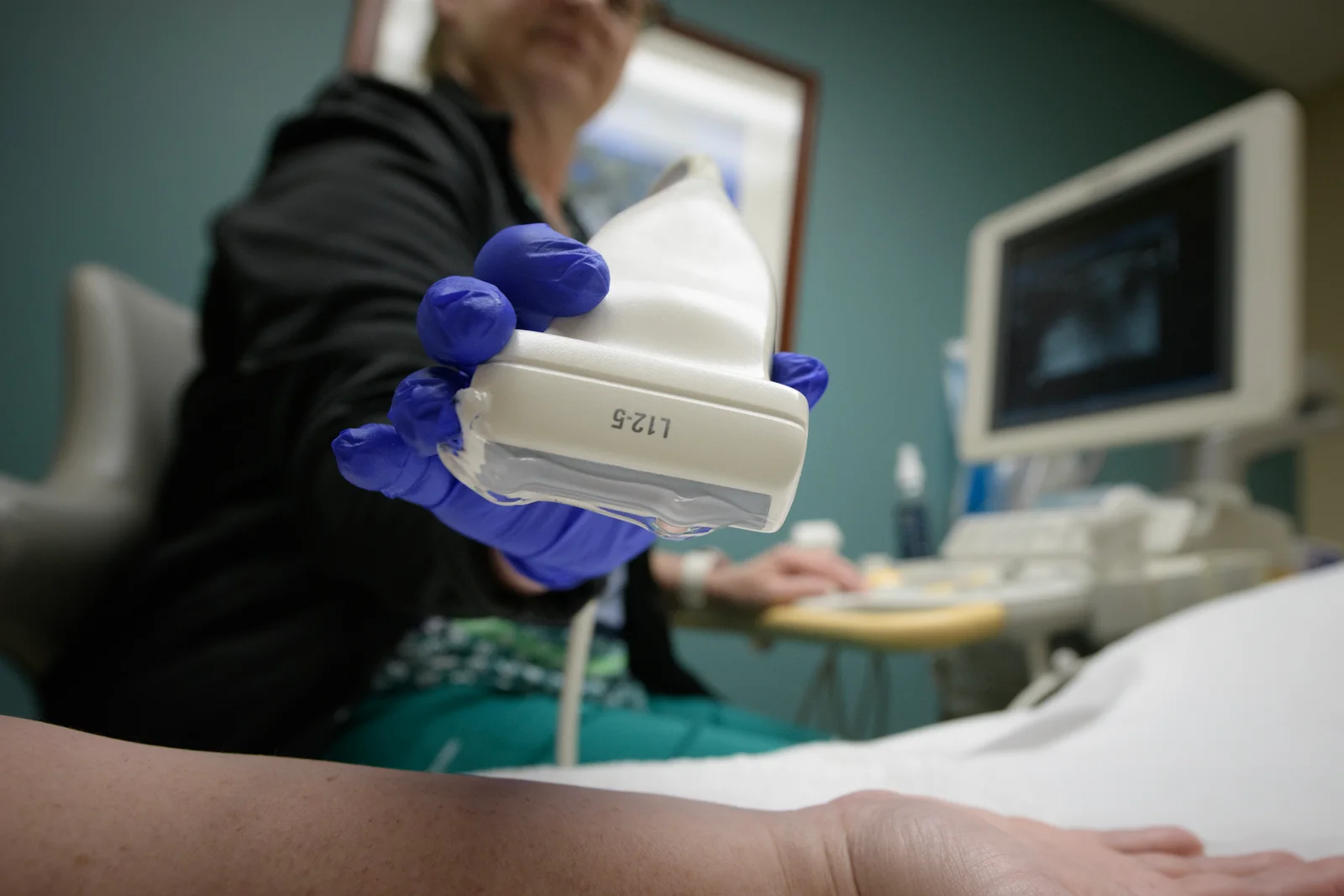
Explain the exam, ask about relevant history, apply gel and position the patient so the area is reachable.

Slide the transducer while watching the live image, adjusting settings, pressure, position and angle for a clearer view.
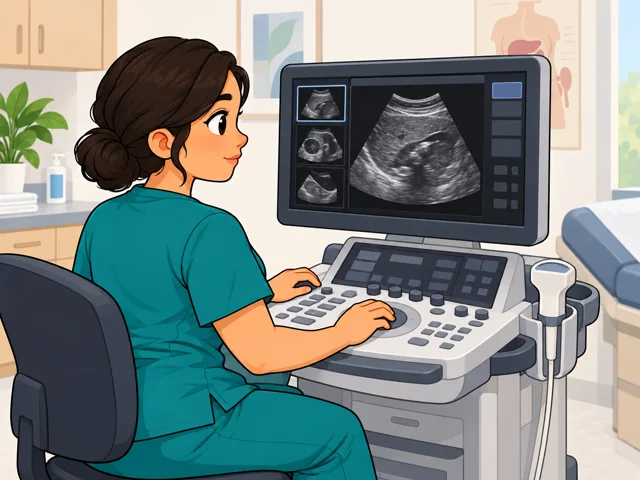
Judge images for quality and coverage, pick keepers and decide if the exam needs another view or widening.
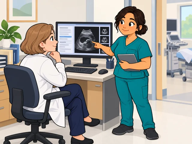
Save usable images, update the record and give the physician a spoken or written summary of technical findings.
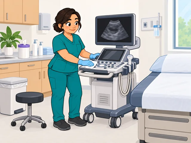
Clean and inspect the equipment, lay out supplies and flag any problem that could disrupt the following exam.
United States
United States · May 2025
United States · 2025–2035
Quick orientation
An associate degree is the typical entry-level education. Postsecondary certificates and bachelor's programs are also available, and programs usually include supervised clinical work.
National median annual wage for diagnostic medical sonographers; this is not a starting wage or a promise of earnings.
National employment is projected to grow 14%, with about 6,000 openings per year on average from growth and replacement needs. Local conditions may differ.
On one FDA-cleared cardiac ultrasound system, machine-learning software can place a subset of 2D and Doppler measurements for the user to edit, accept, or reject.
The sonographer still acquires the views, checks image quality and coverage, reviews the proposed measurements, cares for the patient, and remains responsible for sound clinical judgment.
This feature is specific to one cleared cardiac-ultrasound system, not every workplace. How widely employers use it and whether tools like it affect staffing remain uncertain.
Pay in context
National and industry medians give you two benchmarks for comparing an actual offer.
BLS reported a $96,590 annual median wage for diagnostic medical sonographers. The median hourly wage was $46.44.
In May 2025, the 10th-percentile annual wage was $67,820 and the 90th-percentile wage was $129,370. These are wage percentiles, not starting and senior salaries.
The median was $97,390 in hospitals and $127,940 in outpatient care centers. These are industry-wide figures, not matched offers for workers with the same experience and schedule.
Two common patterns are a sonography degree and a certificate for people with prior healthcare preparation. The American Registry for Diagnostic Medical Sonography sets pathway-specific education, supervised-clinical-experience, and documentation rules for exam eligibility; state licensing and employer requirements may add other steps.
After entry
This starts where the entry routes leave off: early work, growing capability, and advancement or specialization.
In your first role, learn the facility's exam protocols, record accurate histories, produce complete studies, care for patients, and give physicians a clear technical summary.
Strengthen your ability to position patients, adjust equipment, recognize missing views, and adapt an examination without losing track of patient comfort.
Develop depth in a specialty and, when useful for the role, pursue its matching credential. The Registered Diagnostic Medical Sonographer credential requires a Sonography Principles and Instrumentation exam, a specialty exam, and applicable prerequisites.
Hospitals employed 58% of sonographers in 2025. Work may include examinations at a patient's bedside, and facilities that stay open continuously may use evening, weekend, or overnight shifts.
Physicians' offices employed 21% in 2025. The patient group and scan mix may follow the practice's specialty, while the work remains close-contact and equipment-based.
Medical and diagnostic laboratories employed 11% and outpatient care centers employed 5% in 2025. These are dedicated testing settings outside physician offices and inpatient units; case mix and schedules vary by facility.
Try the work pattern
Watch a public ultrasound demonstration from a sonography school or healthcare organization. Note each change in probe position, angle, pressure, patient position, or machine setting—and what the operator says to the patient. This can reveal whether precise hand work, live screen reading, and close patient contact hold your attention.
Stay safe: Use a public demonstration only. Do not scan yourself or another person, and do not try to interpret an ultrasound image.